Deoxyribonucleic Acid (DNA) Tutorial
If your browser/OS combination is Java capable, you will get snappier performance if you use Java. However, the HTML5 version works on more devices.
Overview
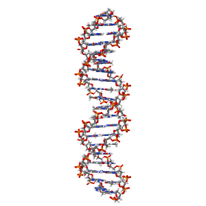
You should be able to view the molecular model of a DNA molecule on the right. This is a simplified model structure of B-form DNA as determined by
studying DNA fibres. You can
the model an anytime.
The original view shows B-DNA with the atoms coloured as follows; Carbon - grey, Oxygen - red,
Nitrogen - blue, Hydrogen - white and phosphorous - orange. The model is displayed in the wireframe form to show the bonds between atoms.
To manipulate the model press and hold the left hand mouse button and drag to obtain the desired view. To zoom in or out, drag the mouse while pressing the
shift key. To alter the presentation of the image or stop the rotation click with the right hand mouse button and select one of the options.
You may prefer to view the wireframe model showing.
A more realistic representation of DNA is the model where the size of each molecule is represented by its bonding properties and van der Waals radius. You may prefer to view the spacefill model showing.
DNA Stucture
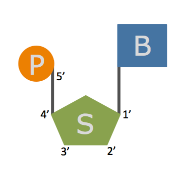
DNA is a long polymer made my joining together similar monomers, called nucleotides, into a long unbranched chain. Above is a cartoon
representation of a nucleotide, it comprises a nitrogenous base (B), the sugar deoxyribose (S) and a phosphate residue (P). The carbon atoms within the
deoxyribose are numbered 1’ to 5’.
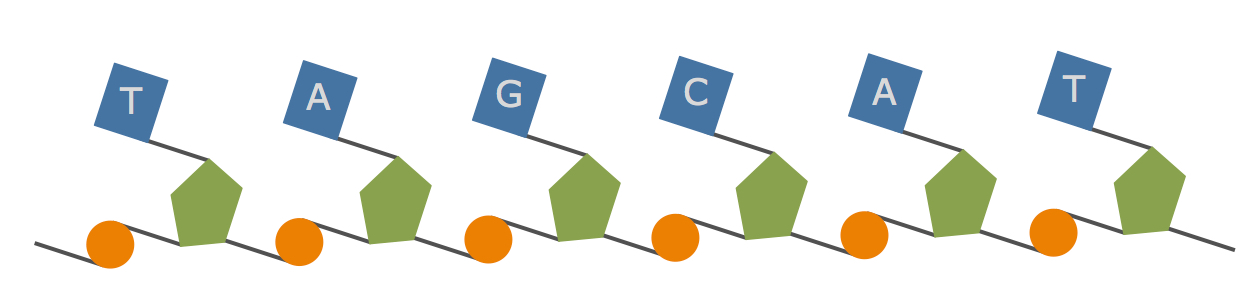
In the cartoon above you will see that the ‘backbone’ of the chain is formed by
alternating phosphate and deoxyribose residues; each deoxyribose will be linked to one phosphate via it’s 3’ carbon and the other phosphate
via it’s 5’ carbon, so a linear DNA chain will have a 5’ end and a 3’ end, this gives each chain a sense of direction. Each nucleotide will contain one
of four possible bases; adenine, guanine, cytosine and thymine. The information in DNA is therefore coded by the sequence of bases.
Within the cell, DNA is double-stranded; two DNA chains pair to form the Watson-Crick double helix structure. The two chains are coiled around a common axis forming a right handed helix. The two chains run in opposite direction to each other, one running 5' to 3' and the other going 3' to 5'. In this one chains is coloured red and the other blue, the 5' end of each chain is darker than the 3' end highlighting that the two strands run in opposite directions.
The and are on the outside of the helix, whereas the are on the inside.
Each base on one strand pairs with a base on the other strand; Adenine (A) always pairs with Thymine (T) and Guanine (G) always pairs with Cytosine (C). between the base pairs holds the two strands together. In the sugar and phosphates are coloured green, whereas the bases are coloured yellow; you can see that the sugar-phosphate backbone has a uniform stucture.
If we and highlight one base (red) and one sugar residue (blue) within the rest of the DNA (white) you can clearly see that the planes of the bases are perpendicular to the helix axis and the plane of the deoxyribose is nearly at a right angle to that of the base.
The arrangement of the two chains results in DNA containing two grooves, ; in this representation atoms facing out into the major groove are coloured pink, whereas atoms facing out into the minor groove are coloured blue; all other atoms are coloured white.
Quick Links